Call us now :08045815486

 Send Inquiry
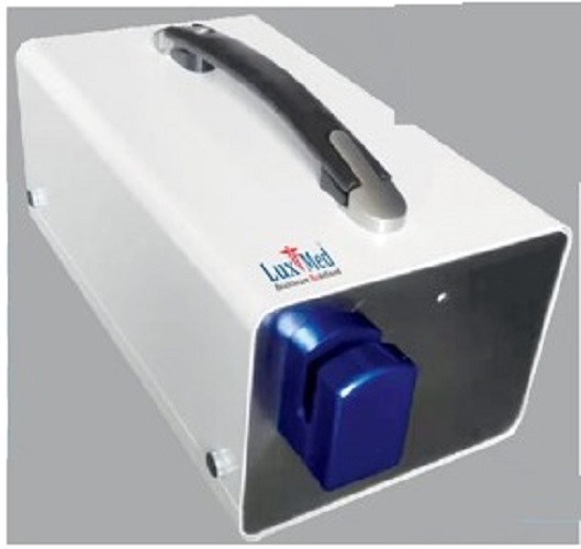
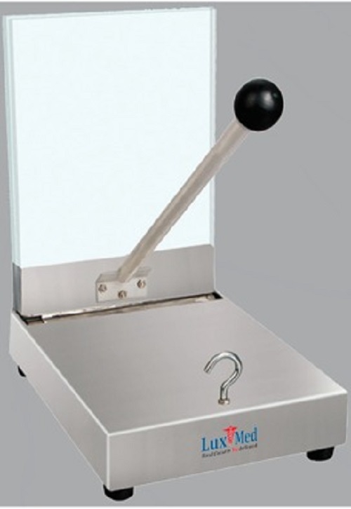
Histology Lab Equipments
Price 150000.0 INR/ Number
Histology Lab Equipments Trade Information
- Minimum Order Quantity
- 1 Number
- FOB Port
- Ambala
- Payment Terms
- Cash in Advance (CID)
- Supply Ability
- 1 Number Per Week
- Delivery Time
- 2 Week
- Sample Available
- Yes
- Sample Policy
- If order is confirmed we will reimburse the sample cost
- Packaging Details
- Wooden Packaging
- Main Export Market(s)
- Asia
- Main Domestic Market
- All India
- Certifications
- CE, ISO , MSME and Startup India
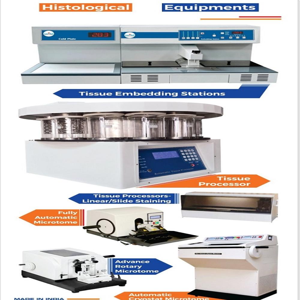
About Histology Lab Equipments
LuxMed Histology lab equipment is usedto prepare and examine tissue samples under a microscope, aiding in disease diagnosis and research of cellular structures.Key instruments include microtomes for sectioning, tissue processors for automated tissue preparation, cryostats for frozen tissue sectioning, and microscopes for observation.These tools are essential for various applications, including diagnostic pathology, research and development, and education.
Specific Equipment and their Uses:
- Microtomes:
These instruments cut thin sections (2-10 micrometers) of embedded tissue, allowing for microscopic examination.There are manual and motorized microtomes, with adjustable cutting thicknesses and feeders.
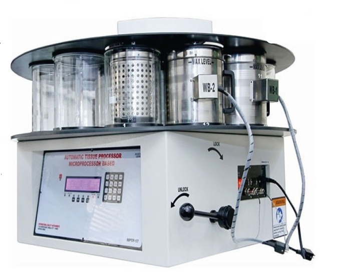
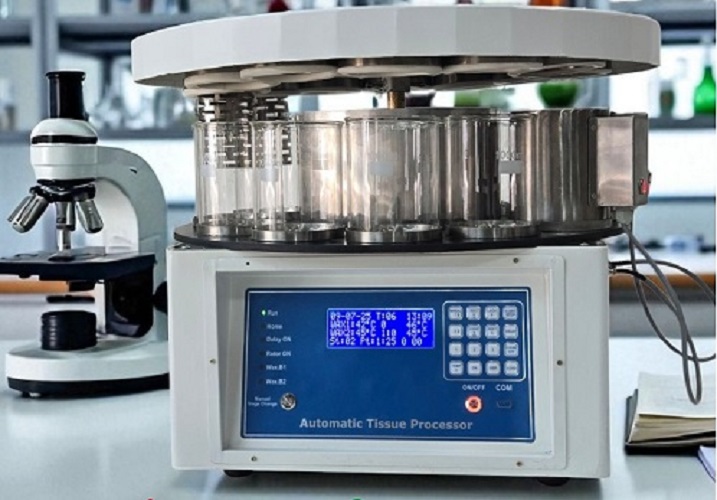
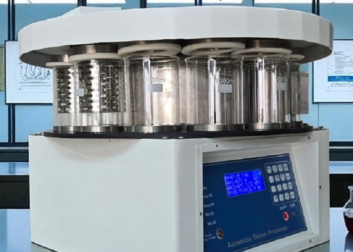
- Tissue Processors:
These machines automate the dehydration, clearing, and infiltration of tissue samples, preparing them for embedding in paraffin.
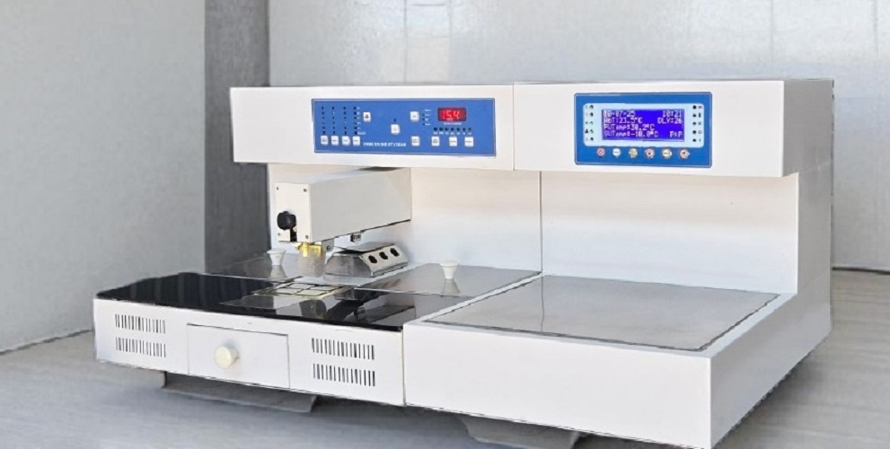
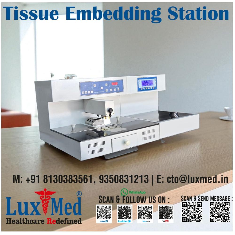
- Embedding Centers:
They are used to embed tissue samples in paraffin wax, creating blocks that can be sectioned by a microtome.
- Cryostats:
Specialized instruments that rapidly freeze tissue samples, enabling sectioning without embedding.This is useful for examining fresh or frozen tissue samples.
- Microscopes:
These are fundamental for viewing the prepared tissue sections, allowing researchers and pathologists to analyze cellular structures and tissue morphology.
- Slide Stainers:
Automated machines that apply stains to tissue sections, highlighting specific cellular components and structures.
- Immunostainers:
Automated machines for immunohistochemistry, which allows for the detection of specific proteins or antigens in tissue sections using antibodies.
- Tissue Flotation Baths and Warmers:
Used to flatten and dry tissue sections on microscope slides.
- Centrifuges:
Used to separate components of tissue samples by centrifugal force, aiding in sample preparation and analysis.
- Slide Storage Units:
Used to store microscope slides, protecting them from contamination and damage.
Tell us about your requirement

Price:
Quantity
Select Unit
- 50
- 100
- 200
- 250
- 500
- 1000+
Additional detail
Mobile number
Email

More Products in Blood Bank Equipments Category
Our Products


B 232, Ground Floor, Khasra No. 832, Chattarpur Extension,New Delhi - 110074, India

Mr Suraj Kumar
(CTO)
Mobile :08045815486
 Send Inquiry
 Send SMS
Developed and Managed by Infocom Network Private Limited.
